
- Products
- Catalogs
- News & Trends
- Exhibitions
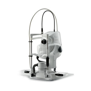
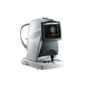
OCT ophthalmoscope Duo™2non-mydriatic retinal cameraretinal autofluorescence imagingtabletop

Add to favorites
Compare this product
Characteristics
- Type of instrument
- OCT ophthalmoscope, non-mydriatic retinal camera
- Examination
- retinal autofluorescence imaging
- Ergonomics
- tabletop
Description
Fundus image acquisition with macula and disc capture in one image on OCT
Combined diagnosis of macular and disc pathologies
Denoising technique with deep learning
Quick acquisition of high definition B-scan images from a single-frame image
Fundus autofluorescence (FAF)
Detailed Information
Wide area scan (12 x 9 mm)
Wide area normative database (macula: 9 x 9 mm, disc: 6 x 6 mm)
A 12 x 9 mm wide area image can be acquired. The retina map captures both the macula and disc in a single shot.
The normative database provides a wide area color-coded map comparing the patient’s macular thickness to a population of normal eyes.
Denoising using deep learning
A new image enhancement technique using deep learning automatically displays a denoised image once B-scan acquisition is complete. With deep learning of a large data set of images averaged from 120 images, this denoising technique provides high definition images comparable to a multiple-image-averaging technique. The denoising function generates high definition images from a single frame while decreasing image acquisition time and increasing patient comfort.
Fundus autofluorescence (FAF)
The FAF function is an advanced screening feature that allows non-invasive evaluation of the RPE without contrast dye.
*Available for the FAF model
*Images courtesy of Kariya Toyota General Hospital
VIDEO
Catalogs
No catalogs are available for this product.
See all of NIDEK‘s catalogsRelated Searches
- Ultrasound system
- B/W ultrasound system
- Tabletop laser
- Nd:YAG laser
- On-platform ultrasound system
- Fixed ophthalmic examination
- Tabletop ophthalmic examination instrument
- Touchscreen ultrasound system
- Ophthalmic biomicroscope
- Hand-held ophthalmic examination instrument
- Table ophthalmic biomicroscope
- Nanosecond laser
- Ophthalmoscope
- Ophthalmic laser
- Refractometer ophthalmic examination
- Automatic refractometer
- Keratometer ophthalmic examination
- Automatic optical lens processing system
- Automatic keratometer
- Tonometer
*Prices are pre-tax. They exclude delivery charges and customs duties and do not include additional charges for installation or activation options. Prices are indicative only and may vary by country, with changes to the cost of raw materials and exchange rates.


