
- Products
- Catalogs
- News & Trends
- Exhibitions
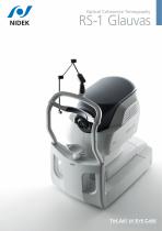
OCT ophthalmoscope RS-1tabletop

Add to favorites
Compare this product
Characteristics
- Type of instrument
- OCT ophthalmoscope
- Ergonomics
- tabletop
Description
250,000 A-scans/s high-speed imaging
Wide, deep, high-resolution imaging
Effortless operation and interpretation
Advanced analytics
The incorporation of 250,000 A-scans/s accelerates your workflow by reducing capture time. The high-speed imaging also addresses patient fixation errors thus contributing to greater image clarity and patient comfort.
Wide, deep, high-resolution imaging
With RS-1 Glauvas, a single B-scan image clearly presents the area from the optic nerve head to the temporal vascular arcade, and the 4.2 mm depth B-scan imaging readily captures the oblate retinal shape of myopic eyes. Improvements in AngioScan OCT-Angiography include wider and clearer images for assessing chorioretinal microvasculature.
Effortless operation and interpretation
Easy image capture with automated functions
The auto alignment and auto switch functions allow anyone to effortlessly capture images. Operators need to only adjust the chinrest height and click Optimize and Capture.
Advanced analytics
Glaucoma analysis in myopia
The long axial length normative database*1 presents analysis with axial length compensation, allowing for a more accurate glaucoma assessment in patients with axial myopia. The OCT Viewer automatically switches to this database as required, by using the axial length*2 which is a parameter for scan width correction.
Less false positives with deep learning segmentation (DL segmentation)
The accuracy of segmentation affects the outcomes of glaucoma analysis.
VIDEO
Catalogs
RS-1 Glauvas
12 Pages
Related Searches
- Ultrasound system
- B/W ultrasound system
- Tabletop laser
- Nd:YAG laser
- On-platform ultrasound system
- Fixed ophthalmic examination
- Tabletop ophthalmic examination instrument
- Touchscreen ultrasound system
- Ophthalmic biomicroscope
- Hand-held ophthalmic examination instrument
- Table ophthalmic biomicroscope
- Nanosecond laser
- Ophthalmoscope
- Ophthalmic laser
- Refractometer ophthalmic examination
- Automatic refractometer
- Keratometer ophthalmic examination
- Automatic optical lens processing system
- Automatic keratometer
- Tonometer
*Prices are pre-tax. They exclude delivery charges and customs duties and do not include additional charges for installation or activation options. Prices are indicative only and may vary by country, with changes to the cost of raw materials and exchange rates.

