
- Products
- Catalogs
- News & Trends
- Exhibitions
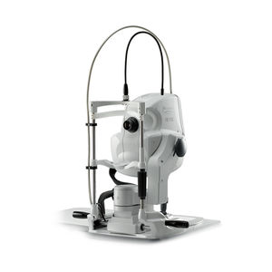
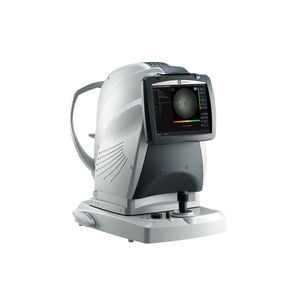
Optical ophthalmic biometer AL-Scanultrasound biometertabletop

Add to favorites
Compare this product
fo_shop_gate_exact_title
Characteristics
- Type of instrument
- optical biometer, ultrasound biometer
- Ergonomics
- tabletop
Description
3D auto tracking and auto shot
Anterior segment observation with Scheimpflug imaging and double mire ring keratometry
Optional built-in ultrasound biometer
IOL power calculation and IOL constants optimization
Additional features with AL-Scan Viewer for NAVIS-EX
Detailed Information
6 clinical parameters in 10 seconds
In 10 seconds, six values for cataract surgery are measured:
- Axial length
- Corneal curvature radius
- Anterior chamber depth
- Central corneal thickness
- White-to-white distance
- Pupil size
3D auto tracking and auto shot
The 3D auto tracking follows eye movements along the X-Y-Z directions to ensure accurate alignment of the eye. Once correct alignment is completed, the auto shot immediately captures the image and data.
Anterior segment observation with Scheimpflug imaging and double mire ring keratometry
The AL-Scan provides sectional lens image, pupil image, and reflected image of double mire rings projected onto the cornea.
Optional built-in ultrasound biometer
In cases where the optical biometer cannot measure an eye with an extremely dense cataract, the AL-Scan provides an optional built-in ultrasound biometer, allowing measurement of virtually any cataractous eye with a combined model. The AL-Scan requires no connection with an external ultrasound unit.
IOL power calculation and IOL constants optimization
The IOL power is automatically calculated after measurement. Calculation of a personalized IOL constant improves postoperative accuracy.
VIDEO
Catalogs
No catalogs are available for this product.
See all of NIDEK‘s catalogsRelated Searches
- Ultrasound system
- B/W ultrasound system
- Tabletop laser
- Nd:YAG laser
- On-platform ultrasound system
- Fixed ophthalmic examination
- Tabletop ophthalmic examination instrument
- Touchscreen ultrasound system
- Ophthalmic biomicroscope
- Hand-held ophthalmic examination instrument
- Table ophthalmic biomicroscope
- Nanosecond laser
- Ophthalmoscope
- Ophthalmic laser
- Refractometer ophthalmic examination
- Automatic refractometer
- Keratometer ophthalmic examination
- Automatic optical lens processing system
- Automatic keratometer
- Tonometer
*Prices are pre-tax. They exclude delivery charges and customs duties and do not include additional charges for installation or activation options. Prices are indicative only and may vary by country, with changes to the cost of raw materials and exchange rates.


