
- Products
- Catalogs
- News & Trends
- Exhibitions
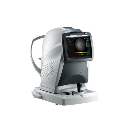
Ophthalmic microperimeter MP-3non-mydriatic retinal camerastatic perimetrytabletop

Add to favorites
Compare this product
Characteristics
- Type of instrument
- micro ophthalmic perimeter, non-mydriatic retinal camera
- Examination
- static perimetry
- Ergonomics
- tabletop
Description
Microperimetry with a wide measurement range
Fixation test with a precise tracking system
High resolution non-mydriatic fundus camera
Feedback exam for visual rehabilitation
Scotopic microperimetry
Auto tracking and auto alignment
Detailed Information
Wide measurement range
The MP-3 has a wider range of stimulus intensity, from 0 to 34 dB, compared to the MP-1. The MP-3 measures perimetric threshold values, even for normal eyes. A maximum stimulus luminance of 10,000 asb* allows evaluation of low-sensitivity.
*In accordance with ISO12866 measurement methods
Fixation test
Precise tracking system
The MP-3 can measure fixation and determine the preferred retinal locus, simply by having the patient fixate on a target Constant tracking of the eye during microperimetry allows evaluation of fixation in patients with central visual field defects and determines whether fixation improves after treatment.
Retinography
High resolution non-mydriatic fundus camera
An easy to use 12-megapixel fundus camera is incorporated into the MP-3 and acquires high resolution images of retinal pathology.
Feedback exam for visual rehabilitation
The visual rehabilitation mode trains low-vision patients who have lost foveal fixation to relocate their preferred retinal locus (PRL) to a different region, called the trained retinal locus (TRL). The TRL is predetermined by a physician, and fixation rehabilitation allows the patient better functional vision (i.e. reading speed) due to increased fixation stability and visual outcomes.
Active flickering pattern stimulation and cheery music create an effective and pleasant training experience for the patient.
VIDEO
Catalogs
Related Searches
- Ultrasound system
- B/W ultrasound system
- Tabletop laser
- Nd:YAG laser
- On-platform ultrasound system
- Fixed ophthalmic examination
- Tabletop ophthalmic examination instrument
- Touchscreen ultrasound system
- Ophthalmic biomicroscope
- Hand-held ophthalmic examination instrument
- Table ophthalmic biomicroscope
- Nanosecond laser
- Ophthalmoscope
- Ophthalmic laser
- Refractometer ophthalmic examination
- Automatic refractometer
- Keratometer ophthalmic examination
- Automatic optical lens processing system
- Automatic keratometer
- Tonometer
*Prices are pre-tax. They exclude delivery charges and customs duties and do not include additional charges for installation or activation options. Prices are indicative only and may vary by country, with changes to the cost of raw materials and exchange rates.


