
- Products
- Catalogs
- News & Trends
- Exhibitions
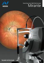
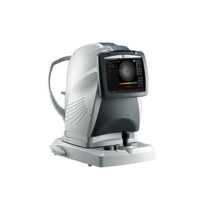
OCT ophthalmoscope MiranteSLO ophthalmoscopetabletop

Add to favorites
Compare this product
Characteristics
- Type of instrument
- OCT ophthalmoscope, SLO ophthalmoscope
- Ergonomics
- tabletop
Description
The ultimate multimodal imaging platform
- Color / FA / ICG / Blue-FAF / Green-FAF / Retro mode
- OCT / OCT-Angiography
Ultra wide field x ultra HD image
Unsurpassed color
Dynamic/Simultaneous FA and ICG
Unique Retro mode
HD wide area OCT
Detailed Information
The ultimate multimodal imaging platform
For the SLO/OCT model
- Color / FA / ICG / Blue-FAF / Green-FAF / Retro mode
- OCT / OCT-Angiography*
For the SLO model
- Color / FA* / ICG* / Blue-FAF /Green-FAF / Retro mode
Streamlined combination capture
The Combo image capture allows sequential capture of images with the preset combination of image capture settings for each specified disease.
Ultra wide field x ultra HD image *
163° ultra wide field image
The clear image of the entire 163° field of view** enables detailed evaluation of pathologies from the fovea to the extreme periphery.
* Ultra wide field imaging is available with the optional wide-field adapter.
** Measured from the center of the eye
Ultra 4K HD and averaging function for unparalleled clarity
4,096 x 4,096 pixel imaging captures every detail of the retina and choroid. Additionally, zooming in allows high magnification, clear visualization of subtle changes in pathology, and resolution of the fine details of capillaries.
The FlexTrack algorithm corrects image distortion due to unstable fixation and enhances averaging quality.
Unsurpassed color
Three separate RGB detectors simultaneously scan different depths of retina with red, green, and blue wavelengths. A color histogram is available for fine adjustment based on pathology or practitioner preference.
VIDEO
Catalogs
Related Searches
- Ultrasound system
- B/W ultrasound system
- Tabletop laser
- Nd:YAG laser
- On-platform ultrasound system
- Fixed ophthalmic examination
- Tabletop ophthalmic examination instrument
- Touchscreen ultrasound system
- Ophthalmic biomicroscope
- Hand-held ophthalmic examination instrument
- Table ophthalmic biomicroscope
- Nanosecond laser
- Ophthalmoscope
- Ophthalmic laser
- Refractometer ophthalmic examination
- Automatic refractometer
- Keratometer ophthalmic examination
- Automatic optical lens processing system
- Automatic keratometer
- Tonometer
*Prices are pre-tax. They exclude delivery charges and customs duties and do not include additional charges for installation or activation options. Prices are indicative only and may vary by country, with changes to the cost of raw materials and exchange rates.


