- Primary care
- General practice
- Eye anatomical model
- Ocular Instruments
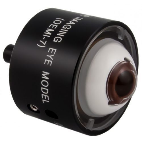
Eye anatomical model OEMI-7for health educationmedical imagingtraining
Add to favorites
Compare this product
Characteristics
- Area of the body
- eye
- Application
- for health education
- Procedure
- training, medical imaging
- Other characteristics
- kit
Description
The most realistic eye model available for Ocular fundus imaging. The unique design incorporates an anterior chamber, crystalline lens, and fundus. Model provides superior demonstration and training of common ophthalmic imaging devices. This eye model incorporates many useful features not available in other eye models, including a retinal detachment showing an elevated retina, a foreign body, optic disc, and blood vessels. In addition, fluorescent features within the eye allow simulated fluorescein imaging. A line at the 180° meridian designates the region of the equator. A peg on the bottom of the model fits into the Ocular Eye Model Bracket (OEMB1) which can be attached to the vertical post of the slit lamp chin rest.
The Ocular Refill Kit (OEMI-KIT) is required for maintenance
Catalogs
Ocular Imaging Eye Model
2 Pages
*Prices are pre-tax. They exclude delivery charges and customs duties and do not include additional charges for installation or activation options. Prices are indicative only and may vary by country, with changes to the cost of raw materials and exchange rates.


