- Products
- Analysis software module
- Optopol Technology
- Products
- Catalogs
- News & Trends
- Exhibitions
Analysis software module T-OCT™captureophthalmologycornea
Add to favorites
Compare this product
Characteristics
- Function
- analysis, capture
- Applications
- ophthalmology
- Area of the body
- cornea
Description
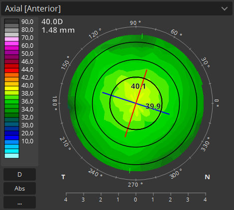
Topography OCT module provides the analysis of both surfaces based on Corneal Curvature, Dioptric power, Elevation and Real power analysis based on both surfaces and local cornea thickness (Ray tracing).
T-OCT™ is a pioneering way to provide detailed corneal Curvature maps by using posterior dedicated OCT. Anterior, Posterior surface and Corneal Thickness allow to provide the True Net Curvature information. With Net power, the precise understading of the patient’s corneal condition comes easily and is free of errors associated with modelling of posterior surface of the cornea. SOCT T-OCT module provides Axial maps, Tangential maps, Total Power map, Height maps, Epithelium and Corneal thickness maps.
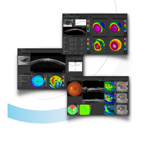
Corneal topography module clearly shows the changes in the cornea on the difference map view. Customize your favoured view by selecting a variety of available maps and display options. Fully Automatic module capture with examination time of up to 0.3 sec makes testing quick and easy.
Topography module provides:
Full featured Corneal mapping of Anterior, Posterior and Real
Precise Astigmatism Display Option (SimK: Anterior, Posterior, Real, Meridian and Emi-Meridian ø 3, 5, 7 mm zones)
Keratoconus Screening
Easily detect and classify keratoconus with the Keratoconus classifier. Classification based on KPI, SAI, DSI, OSI and CSI. In the early stages of keratoconus the results can be complemented by Epithelium and Pachymetry maps.
Catalogs
*Prices are pre-tax. They exclude delivery charges and customs duties and do not include additional charges for installation or activation options. Prices are indicative only and may vary by country, with changes to the cost of raw materials and exchange rates.


