- Products
- Management software module
- Optopol Technology
- Products
- Catalogs
- News & Trends
- Exhibitions
Measurement software module B-OCT® evaluationmanagementscan
Add to favorites
Compare this product
Characteristics
- Function
- measurement, evaluation, management, scan
- Applications
- ophthalmology
- Area of the body
- retina, cornea, pupil
Description
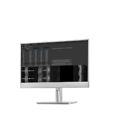
B-OCT™ Innovative method of using the posterior OCT device to measure ocular structure along eye axis. OCT Biometry provides a complete set of Biometry parameters: Axial Length (AL), Central Cornea Thickness (CCT), Anterior Chamber Depth (ACD), Lens Thickness (LT), White to White (WTW), Pupil (P).
Overview
B-OCT® enables measurement of ocular axial dimensions using a conventional OCT system. During the examination, the scanning light beam passes through ocular structures located along the visual axis and the following are identified: the anterior and posterior boundary of the cornea, the anterior and posterior boundary of the lens, as well as the posterior boundary of the retina.
B-OCT® provides a complete set of Biometry parameters:
Axial Length (AL)
Cornea thickness (CTT)
Anterior chamber depth (ACD)
Lens thickness (LT)
White to White (WTW)
Pupil (P)
Verify your measurement visually
All measurement callipers are shown on all boundaries of OCT image provided by REVO. Now, you can visually verify, identify and if need be, make corrections as to which structure of the eye has been measured. With a simple cursor shift it is possible to precisely set boundaries for every difficult patient. From now on you can eliminate the common uncertainty as to how the optical biometer classifies the boundaries in non-typical patients.
3 mm scan allows precise evaluation of the Retina condition,
5 µm resolution gives the highest level of details.
Catalogs
REVO NX 130
16 Pages
REVO 60
8 Pages
*Prices are pre-tax. They exclude delivery charges and customs duties and do not include additional charges for installation or activation options. Prices are indicative only and may vary by country, with changes to the cost of raw materials and exchange rates.


