- Products
- Catalogs
- News & Trends
- Exhibitions
Analysis software SOCT 11.5scantrackingwith artificial intelligence
Add to favorites
Compare this product
Characteristics
- Function
- analysis, scan, tracking, with artificial intelligence
- Applications
- ophthalmology
- Area of the body
- cornea, retina
- Type
- 3D
Description
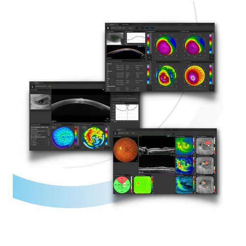
The innovative AI technology provides precise and reliable algorithms for analyzing the epithelium and stroma layers.
AI Lens segmentation algorithms. High-precision algorithms for challenging B-OCT cases.
3D Widefield analysis. Fast overview of the condition of the retinal and ganglion cells layers.
Fundus image processing tools. Enable the clinician to extract hidden information even in the case of patients with cataracts.
Full Range Both eyes report for Radial and Line scans
Exporting presented exam results to .csv files
Retina 21 Raster scanning program
Extended features in Auto Backup functions
Improvements:
ACCUtrack – new algorithm for hardware eye tracking (only for the REVO FC and the REVO FC 130)
Maximum width of the Retina 3D scan extended to 15×15 mm
Averaging algorithms (better results in Full Range scans)
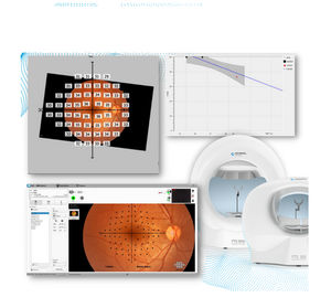
Decreased time of loading perimeter exams in S&F
Added fast access to the Fundus photo preview from an OCT scan
Reduced loading time in the Progression tab
Added 12/4 NFL pie chart data presentation
Catalogs
No catalogs are available for this product.
See all of Optopol Technology‘s catalogs*Prices are pre-tax. They exclude delivery charges and customs duties and do not include additional charges for installation or activation options. Prices are indicative only and may vary by country, with changes to the cost of raw materials and exchange rates.


