- Laboratory
- Laboratory medicine
- 3D viewing software
- Oxford Instruments
Image analysis software Imaris reporting3D viewingmeasurement
Add to favorites
Compare this product
fo_shop_gate_exact_title
Characteristics
- Function
- image analysis, reporting, 3D viewing, measurement, tracking
- Applications
- for digital microscopes, for life sciences applications, for molecular biology
- Area of the body
- blood vessels
- Type
- automated, 3D
Description
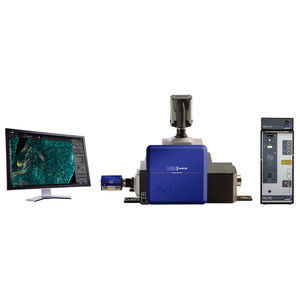
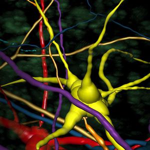
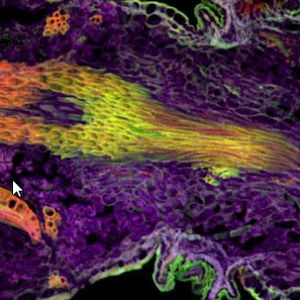
Imaris for Core Facilities is our recommended image analysis software package for microscopy facilities, larger labs and multi-user environments. Imaris is the market-leading visualization and analysis software for widefield, confocal (including spinning disk), light sheet, two-photon, electron microscopy, CLEM, OPT and many other modalities with over 4,000 publications in 2020 alone. Imaris for Core Facilities provides smart detection and visualisation of complex objects, tracing of neurons, blood vessels and other filamentous structures, 3D tracking (including cell division detection), interactive plotting and batch analysis.
3D Microscopy Image Analysis
The power of Imaris for Core Facilities lies in our multiple detection models and the versatility they provide for all users and their research problems. Our 4 models (Spots, Surfaces, Cells and Filaments) can be harnessed to detect and analyse almost all biological samples, including cells, nuclei, nucleoli, bacteria, viruses, organs, neurons, dendritic spines, blood vessels and many more. Using them in combination alongside Imaris’ other tools opens the possibility to analyse dynamics of objects over time as well as their relationships with other objects in the image and to present these data in an elegant and informative fashion.
Quantitative Image Analysis for 3D Microscopy
After detecting and segmenting the image data as objects using one of the 4 models, Imaris calculates a wide range of statistics.
Image Analysis for Everyone
All users of your facility will experience the simplicity of the Imaris software while using our wizard-driven smart workflows and software hints.
Catalogs
Related Searches
- Analysis medical software
- Microscopy
- Compound microscope
- Laboratory microscope
- Tabletop microscope
- Viewer software
- Control software
- Laboratory software
- Reporting software
- CMOS camera
- USB camera
- Automated software
- LED illuminator
- Acquisition software
- Scan software
- Tracking software
- Biological microscope
- Measurement software
- Microscopy camera
- Research microscope
*Prices are pre-tax. They exclude delivery charges and customs duties and do not include additional charges for installation or activation options. Prices are indicative only and may vary by country, with changes to the cost of raw materials and exchange rates.


