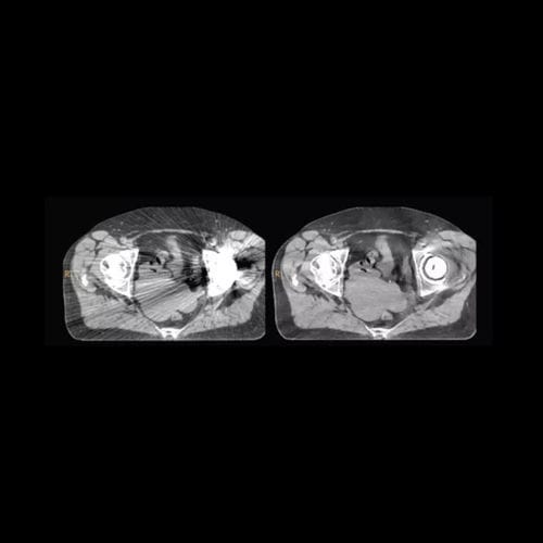
O-MAR helps to improve visualization of anatomy by reducing artifacts related to beam hardening, photon starvation and streaking caused by metal inside the body.
Features
O-MAR improves image quality
O-MAR can be used to isolate the effects of large metal objects in the image data, which results in increased visualization of anatomic structures in areas of the body where the metal implants are present.
Fast and efficient images
O-MAR is applied to the data from a single scan, and the algorithm is an integral part of the scanning protocol, making the process to obtain corrected images fast and efficient.
O-MAR improves contouring and patient marking workflow
Metal from orthopedic implants can cause artifacts in the image data impairing visualization of anatomy, making it very difficult and time-consuming to generate contours of critical structures and target volumes. O-MAR improves visualization of anatomic structures and target volumes for increased workflow in contouring those structures as part of simulation and patient marking.


