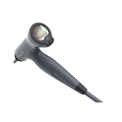
Pediatric video otoscope Tomiscopewith OCT imagingwith integrated video monitorwith speculum
Add to favorites
Compare this product
Characteristics
- Patient type
- pediatric
- Options
- with OCT imaging, with integrated video monitor, with speculum
Description
Real-time middle ear morphology plus eardrum surface video.
Cross-sectional images of the middle ear are shown on the system’s screen, alongside video of the surface of the eardrum. The physician can evaluate the revealing OCT visual images of the middle ear without giving up the more familiar otoscopic view. Images can be saved for later analysis with the click of a button.
Our easy-to-use device is designed to look and handle like the familiar otoscope. OCT images of the middle ear eliminate subjectivity and guesswork for the first time. A nurse-practitioner, physician’s assistant, or technician could perform the exam. This will free up the ENT specialist’s and make lower-cost, objective early screening possible.
*Prices are pre-tax. They exclude delivery charges and customs duties and do not include additional charges for installation or activation options. Prices are indicative only and may vary by country, with changes to the cost of raw materials and exchange rates.

