Dermatology camera RCS-100for endoscopesmonitoringdigital

Add to favorites
Compare this product
Characteristics
- Applications
- dermatology, for endoscopes, monitoring
- Technology
- digital
- Resolution level
- full HD
- Sensor type
- CMOS
- Resolution
- 8 Mpx
- Light source
- LED
- Configuration
- portable
- Other characteristics
- USB, WiFi
- Weight
96 g
(3 oz)
Description
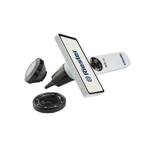
The RCS-100 Wireless Medical Camera System with otoscope lens and LED light provides an enhanced view of the auditory canal, as well as 6 x magnification displayed on the 5” LCD screen.
RCS-100 with otoscope lens can substitute or replace the traditional otoscope, providing the additional benefit of capturing and sharing the digital photograph or video of the ear canal and tympanic membrane. The otoscope lens can be used with a choice of reusable or disposable specula.
Front view of the RCS-100 with otoscope lens.
Otoscope lens.
Disposable specula for otoscope lens.
Side view of the RCS-100 with otoscope lens and specula.
Photographic images of tympanic membranes in routine clinical practice
ENT & Audiology News notes: The ability to capture and record photographic images of tympanic membranes in routine clinical practice has several distinct advantages over hand-drawn representations. Photographs are helpful when explaining unfamiliar concepts to patients and can be a valuable aid when discussing treatment options. Photo documentation is essential for accurately monitoring evolving pathologies, such as tympanic membrane retraction, where subtle changes over time could be easily missed or overlooked when relying on drawings alone. Finally, photographs also provide a precise, contemporaneous record for medico-legal purposes.
Product details
Max object distance 15mm, at max object distance FOC diameter: 15mm
Depth of field scope 10mm
Lighting source Natural light LED
LED colour temperance 4000k
Disposable and reusable specula 2mm (0.08in), 3mm (0.12in), 4mm (0.16in), 5mm (0.2in)
VIDEO
Catalogs
Telemedicine Cart
2 Pages
*Prices are pre-tax. They exclude delivery charges and customs duties and do not include additional charges for installation or activation options. Prices are indicative only and may vary by country, with changes to the cost of raw materials and exchange rates.


