- Products
- Cell imaging software module
- Sartorius Group

- Company
- Products
- Catalogs
- News & Trends
- Exhibitions
Cell imaging software module Incucyte® image analysiscapturetreatment
Add to favorites
Compare this product
Characteristics
- Applications
- cell imaging
- Function
- image analysis, capture, treatment, inspection, navigation
- Type
- automated
Description
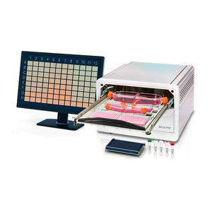
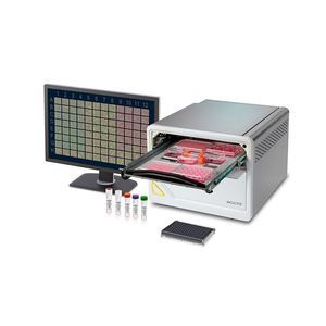
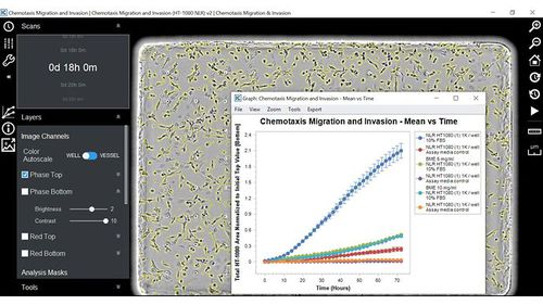
High-definition images and movies enable detailed inspection of morphology and phenotypic treatment effects
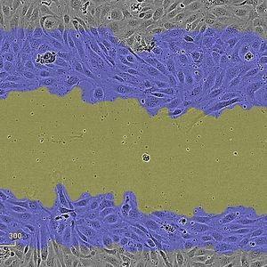
Digitally zoom into any image and visually monitor cells in your experiment over time
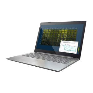
Intuitive image review tools enable quick navigation to data from any well at any time point
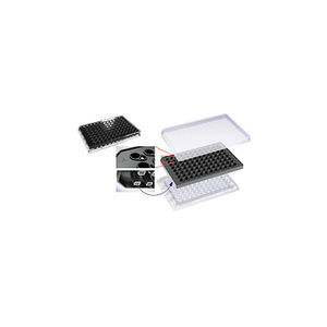
Figure 1. Monitor every cell in your experiment. Capture dynamic migration of cell and digitally zoom into any region of the high-definition images and inspect morphology. Orange circles indicate the location of pores in the Clearview membrane.
User-friendly setup and fully automated image acquisition
Simple user interface for simple experimental set-up
Acquire images over hours or days while cells remain stationary while optics move
Powerful image analysis tools
Quantify cells on the top and bottom surfaces of the Clearview membrane
Review and modify image analysis parameters with ease– you're in control
Quantify cells as they leave the upper surface of the membrane or as they adhere to the lower surface
*Prices are pre-tax. They exclude delivery charges and customs duties and do not include additional charges for installation or activation options. Prices are indicative only and may vary by country, with changes to the cost of raw materials and exchange rates.


