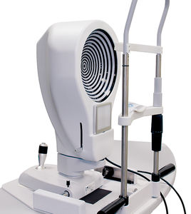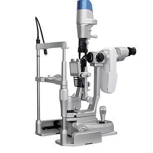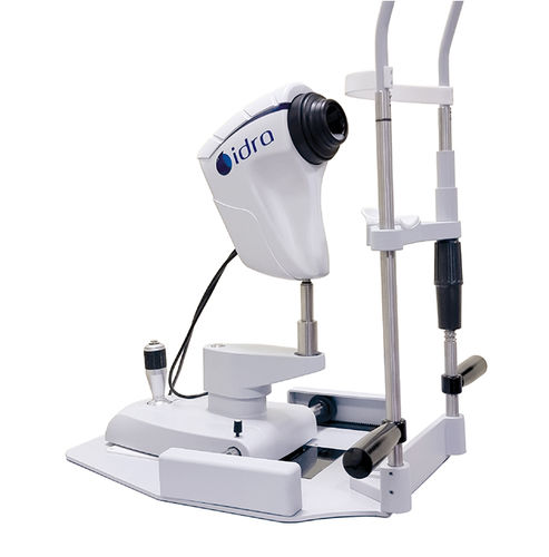
Meibography dry eye diagnosis system IDRAinterferometrytear meniscometry

Add to favorites
Compare this product
Characteristics
- Method
- meibography, tear meniscometry, interferometry
Description
AUTO INTERFEROMETRY
AUTOMATED LIPID LAYER ANALYSIS
The IDRA software analyses lipid layer thickness and allows to understand the functionality of Meibomian Glands.
It is possible to carry out a follow up after MG treatment detecting an Increase in secretion.
AUTO TEAR MENISCUS
Low tear production may resuit in aqueous tear deficiency (ATD) and cause dry eye symptoms.
However, measuring the tear volume is difficult since the methods available nowadays are invasive and irritating.
The analysis of the thickness of the tear meniscus is automatic.
AUTO NIBUT
Thanks to one single video, the physician can gain lots of information:
- Automatic NIBUT
- Average of more than one value
- Graph to understand the trend of tear film stability during the video
- Tear topography that shows ail breaking the tear film during time.
EYE BLINK
Most blinks are spontaneous, occurring regularly with no external stimulus.
However, a reflex blink can occur in response to external stimuli such as a bright light, a sudden loud noise, or an object approaching towards the eyes.
MEIBOGRAPHY
The System automatically analyses the images taken through a sensitive infrared camera (NIR) to locate the Meibomian Glands in a guided way:
- An exam valid both for the upper and the lower eyelids
- Automatic percentage of the Meibomian Gland loss area
3D MEIBOGRAPHY
The revolutionary introduction of the 3D Meibomian Gland imaging gives two big advantages.
Firstly, it enables to confirm the presence of abnormal glands compared to a healthy subject in a 3D view; secondly, it provides a clear image to share with the patients, to help explain the potential reason of their discomfort.
VIDEO
Catalogs
IDRA
4 Pages
Related Searches
- Fixed ophthalmic examination
- Tabletop ophthalmic examination instrument
- Ophthalmic biomicroscope
- Table ophthalmic biomicroscope
- Digital slit lamp
- Corneal topographer
- Vision chart
- Dry eye diagnosis system
- Meibography dry eye diagnosis system
- Tear meniscometry dry eye diagnosis system
- Interferometry dry eye diagnosis system
*Prices are pre-tax. They exclude delivery charges and customs duties and do not include additional charges for installation or activation options. Prices are indicative only and may vary by country, with changes to the cost of raw materials and exchange rates.


