- Medical Imaging
- Radiology
- Medical imaging software
- Shimadzu Europe Medical Systems

- Products
- Catalogs
- News & Trends
- Exhibitions
Medical imaging software SLOT Advanceradiation dose managementpatient dose managementvisualization
Add to favorites
Compare this product
Characteristics
- Applications
- medical imaging, radiation dose management
- Function
- patient dose management, visualization, acquisition, measurement, post-processing
- Area of the body
- spine, leg
Description
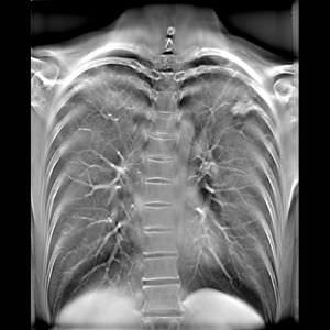
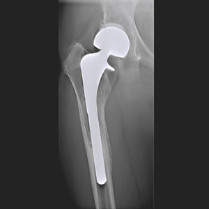
SLOT Advance acquires a series of accurate images of a few centimetres central slit as the imaging chain moves successively along the patient.
Providing super high accuracy measurements SLOT Advance collimates the X-ray beam, exposing just a narrow central slit field without the distortion caused by oblique rays. These central slit images are captured using the best-in-class SONIALVISION R/F table’s super-high resolution Flat Panel Detector (FPD) by moving it in parallel with the X-ray tube. These successive images are reconstructed automatically to create one long image in SONIALVISION G4’s digital imaging unit (DR-300). This simple workflow enables highly accurate measurements without distortion.
Significant reduction of examination time:
Before starting the exposure, the start and end positions of the examination field are simply set. That’s all it takes to obtain proper long-view images using Slot Radiography. All post-processing required to connect, adjust, and display the images on the monitor is done automatically immediately after exposure. SLOT Advance eliminates the time-consuming steps of setting up the cassette and making adjustments, not to mention moving the patient between standing and horizontal positions, all of which are required by conventional CR long-length imaging.
Catalogs
No catalogs are available for this product.
See all of Shimadzu Europe Medical Systems‘s catalogsOther Shimadzu Europe Medical Systems products
Core imaging technologies
Related Searches
- Analysis software
- Radiology software
- Viewer software
- Tablet PC software
- Control software
- Digital radiography system
- Radiography radiography
- Flat panel sensor
- Multipurpose radiography system
- Radiography system
- Radiography system with table
- Automated software
- Diagnostic software
- Acquisition software
- Radiography system with floor-standing bucky
- Portable flat panel sensor
- Treatment software
- Measurement software
- Mobile X-ray unit
- Surgical software
*Prices are pre-tax. They exclude delivery charges and customs duties and do not include additional charges for installation or activation options. Prices are indicative only and may vary by country, with changes to the cost of raw materials and exchange rates.

