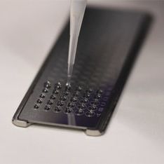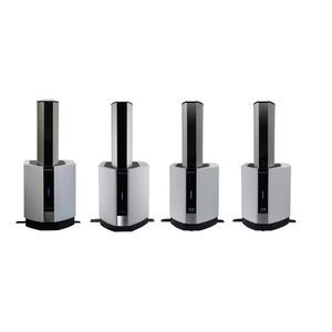
Optical microscope iMScope QTlaboratorybenchtop

Add to favorites
Compare this product
Characteristics
- Type
- optical
- Applications
- laboratory
- Configuration
- benchtop
Description
Next-Generation Mass Spectrometry Imaging Created by iMScope™ QT
Inheriting the concept of a mass spectrometer equipped with an optical microscope from the iMScope series, the iMScope QT is also Shimadzu's flagship model for MS imaging with a LCMS-Q-TOF.
The iMScope QT boasts not only fusion with morphology studies but also excellent speed, sensitivity, and spatial resolution, clearing the way to next-generation mass spectrometry imaging.
Total System for MS Imaging Analysis
Mass spectrometry imaging is performed in three steps: pretreatment, data acquisition, and data analysis.
At each step, the optimal approach accelerates research, while improving the reliability of the results.
Features
Combined Analysis
Fusion of observations from an optical microscope with MS images (exclusive to Shimadzu)
MS images can be obtained flexibly and matched to observation images, either the entire image area or detailed portions of it.
Measurement Results for the Cerebellum with 5 μm Spatial Resolution
Sample :Whole mouse brain
Matrix :9-AA
Measurement region :662×595(393890pix)
Measurement time: :around 2.2 hours
Measurement Results for Whole Brain Sections in Negative Mode
Sample:Whole mouse brain
Matrix :9-AA
Measurement region: :1126×624(702624pix)
Measurement time:around 6 hours
Quantification and Distribution
Obtain qualitative and quantitative information from LC-MS as well as position information from mass spectrometry imaging (MSI) with a single instrument.
The combined system, which can perform LC-MS analysis in addition to MSI analysis, provides both distribution information and quantitative analysis.
VIDEO
Catalogs
No catalogs are available for this product.
See all of Shimadzu‘s catalogsRelated Searches
- Analysis software
- Microscopy
- Compound microscope
- Laboratory microscope
- Tabletop microscope
- Control software
- Laboratory software
- Monitoring software
- Digital microscope
- Zoom microscope
- Compact microscope
- Data analysis software
- Microscope slide
- IR microscope
- 3D microscope
- Small microscopy
- Mass spectrometry software
- Scanning probe microscope
- FT-IR microscope
*Prices are pre-tax. They exclude delivery charges and customs duties and do not include additional charges for installation or activation options. Prices are indicative only and may vary by country, with changes to the cost of raw materials and exchange rates.


