- Company
- Products
- Catalogs
- News & Trends
- Exhibitions
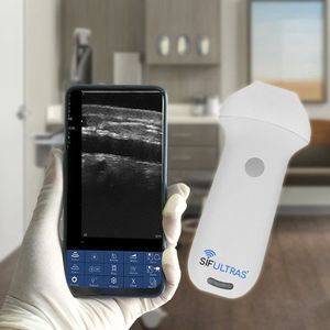
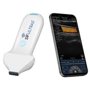
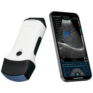
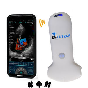
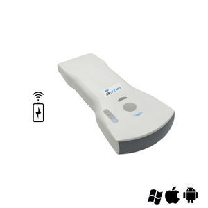
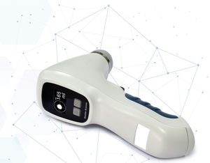
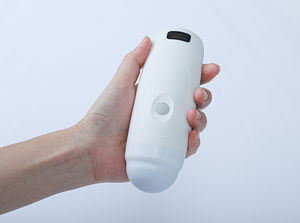
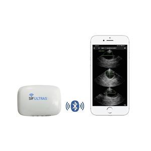
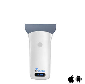
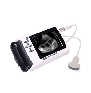
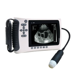
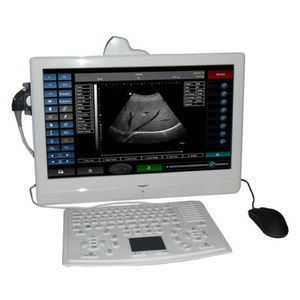
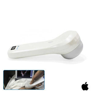
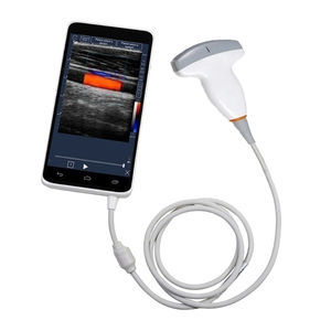
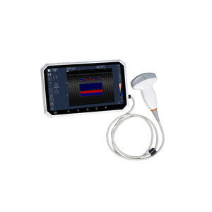
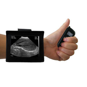
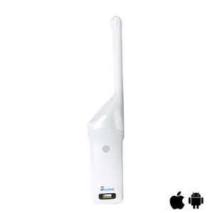
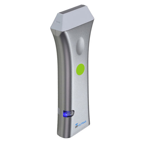
Hand-held ultrasound system SIFULTRAS-5.38for breast ultrasound imagingfor skin ultrasound imagingB/W

Add to favorites
Compare this product
Characteristics
- Ergonomics
- hand-held
- Application
- for breast ultrasound imaging, for skin ultrasound imaging
- Imaging modes
- B/W, color doppler
- Options
- all-in-one probe, wireless probe
- Probe type
- linear-array
- Probe frequency
Max.: 10 MHz
Min.: 5 MHz
- Weight
290 g
(10.23 oz)- Length
38 mm
(1.5 in)- Depth
12 cm
(4.7 in)
Description
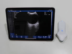
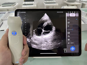
An ultrasound scanner is the third eye of a doctor. The Color 5-10 MHz Wireless Linear Ultrasound Scanner 128 Elements SIFULTRAS-5.38 is a wireless ultrasound scanner. It allows the doctor to make clinical applications anywhere, anytime. The color linear SIFULTRAS-5.38 has 5-10 MHz frequency and 40-120mm.
Also, the SIFULTRAS-5.38 image mode is B, M and color. The B-mode or brightness mode: produced by scanning the transducer beam in a plane as shown in Fig. 1.13. It can be used for both stationary and moving structures such as cardiac valve motion. On the other hand, the M-mode or motion mode: it displays the A-mode signal corresponding to repeated pulses in a separate column of a 2-D image. It is mostly employed in conjunction with ECG for motion of the heart valves.
Consequently, the SIFULTRAS-5.38 is the leading modality in vascular access. For instance, Vascular cannulation: Intravenous (IV) cannulation is a technique in which a cannula is placed inside a vein to provide venous access. Venous access allows sampling of blood, as well as administration of fluids, medications, parenteral nutrition, chemotherapy, and blood products. Further, the SIFULTRAS-5.38 is used for calculation of the speed of blood flow in the vessel, valuation of blood flow in the arteries and veins of the body and to diagnose blood clots in the veins of the arms and legs.
The major advantage of ultrasound is the real-time availability that is particularly important for intraoperative imaging. Ultrasound is used as navigation tool in traditional surgical interventions and in guided biopsies, such as biopsy of breast tumors.
VIDEO
Related Searches
- Ultrasound system
- B/W ultrasound system
- Color doppler ultrasound system
- Multipurpose ultrasound imaging system
- Portable ultrasound system
- Convex-array ultrasound system
- Linear-array ultrasound system
- On-platform ultrasound system
- Hand-held ultrasound system
- Wireless probe ultrasound system
- Touchscreen ultrasound system
- Spectral doppler ultrasound system
- 3D/4D ultrasound system
- Microconvex-array ultrasound system
- Phased-array ultrasound system
- Urology ultrasound imaging system
- Cardiovascular ultrasound imaging system
- Endocavitary ultrasound system
- 15-inch ultrasound system
- Breast ultrasound imaging system
*Prices are pre-tax. They exclude delivery charges and customs duties and do not include additional charges for installation or activation options. Prices are indicative only and may vary by country, with changes to the cost of raw materials and exchange rates.


