- Laboratory
- Laboratory medicine
- Microscope slide scanner
- SigTuple Technologies
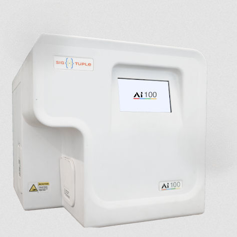
Microscope slide scanner AI100automaticcompact

Add to favorites
Compare this product
Characteristics
- Type of support
- for microscope slides
- Other characteristics
- automatic
- Configuration
- compact
Description
An in-vitro diagnostic device designed to automate manual microscopy in a diagnostic laboratory
It uses robotics and AI to digitize any biological sample on a glass slide to enable AI aided remote review.
One device,
multiple tests!
Can digitize and analyse both blood and urine
Automated,
cell identification!
Backed by visual evidence
Review report from
anywhere & anytime
Web-enabled reports allow WFH for Pathologists!
Image Analysis
Solutions
Benefits
Faster
TAT
Achieve quicker TAT through remote review and reduced review time
Remote Collaboration
Collaborate with colleagues on special cases without being hindered by geographical separation
Improve Patient Outcome
Highly sensitive in finding rarer cells and abnormalities
Reduce Eye Strain and Fatigue
View everything on a large screen. No more peering into a microscope all day
Unlimited Cloud Storage
No more worrying about storage running out. We take care of all your data. Forever!
Quality Check
We ensures sample preparation quality by flagging sub-optimal samples
Predictive Maintenance
We flag anomalies in the system to reduce the probability of any breakdown
Hassle-free Upgrades
Remote upgrades for better performance and new products
Tech Specs
Major Components
Slide or Cartridge holding stage
XYZ motion stage
Optics unit with Turret mechanism unit
Optics digital camera
Display unit
Control unit
Casing
Compatible Optional Analyser Softwares
Sigtuple Shonit- Blood Analyser
Sigtuple Shrava- Urine Analyser
Camera
CMOS sensor
14 MegaPixel resolution
Optics system
Single optical column
40X Objective lens
Optical resolution of approx. 700 lp/mm
VIDEO
Catalogs
Related Searches
*Prices are pre-tax. They exclude delivery charges and customs duties and do not include additional charges for installation or activation options. Prices are indicative only and may vary by country, with changes to the cost of raw materials and exchange rates.


