
- Products
- Catalogs
- News & Trends
- Exhibitions
Ultrasound imaging simulation module for abdominal peripheral interventionanatomyweb-based

Add to favorites
Compare this product
Characteristics
- Procedure
- for ultrasound imaging, for abdominal peripheral intervention
- Application
- anatomy
- Technology
- web-based
Description
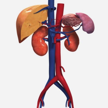
This module teaches you how to prepare for and perform an ultrasound examination of the abdominal vasculature. The interactive simulator provides three different scan scenarios, including normal and pathological cases. It enables students and practitioners to build or refresh knowledge and cognitive skills, and offers a safe online practice environment so you can prepare for the real clinical world. Ideal if you are studying for the American Registry for Diagnostic Medical Sonography (ARDMS) registry exams.
Indications for ultrasound of the abdominal blood vessels
Identify abdominal vascular anatomy on diagrams and sonograms
List signs and symptoms of abdominal vascular disease
Abdominal vascular protocol
Equipment preparation - transducer and preset selection
Patient preparation
Patient positioning
Transducer positions
Scan planes
Identify and obtain sonographic images of the aorta, abdominal aortic branches, and common iliac arteries
Identify and obtain sonographic images of the inferior vena cava and its tributaries
Identify and obtain sonographic images of the portal vein, superior mesenteric vein, inferior mesenteric vein, and splenic vein
Obtain Doppler spectral traces of the aorta and inferior vena cava
Explain and demonstrate the use of breathing techniques to obtain optimal sonographic images of the blood vessels
Differentiate normal and abnormal sonographic appearances of the vascular system
Identify and describe common pathology of the abdominal vasculature
Explain the important ultrasound characteristics when evaluating an abdominal aortic aneurysm
Describe the normal and abnormal Doppler patterns of the vascular structures
VIDEO
Related Searches
*Prices are pre-tax. They exclude delivery charges and customs duties and do not include additional charges for installation or activation options. Prices are indicative only and may vary by country, with changes to the cost of raw materials and exchange rates.


