- Medical Imaging
- Radiation protection, Dosimetry
- CT scan test phantom
- True Phantom Solutions
- Company
- Products
- Catalogs
- News & Trends
- Exhibitions
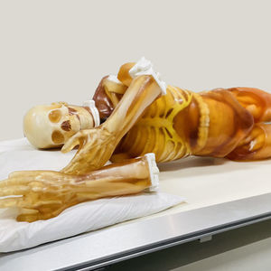
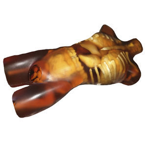
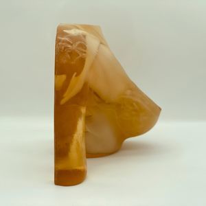
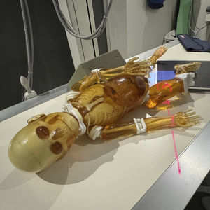
Radiography test phantom SE-A01for CT scanfor MRIspine

Add to favorites
Compare this product
Characteristics
- Type of calibration
- for radiography, for CT scan, for MRI
- Area of the body
- spine
Description
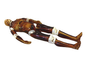
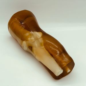
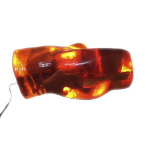
Adult Spine Phantom consists of 25 individual vertebrae assembled on top of each other and encased in a semi-cylindrical-shaped body-mimicking tissue. The vertebrae have a realistic three-layered structure with inner porosity which can be adjusted according to the requirement of the particular project.
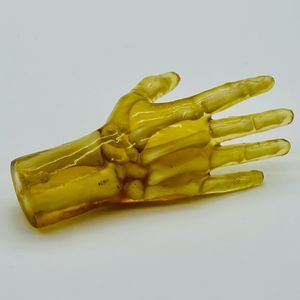
The Phantom is designed based on average human anatomy, and the bones are made out of realistic patented bone material that is suitable for MRI and X-Ray CT applications. It can be used for medical imaging research and treatment planning of various medical procedures. (Upon request, it can be modified for a lumbar or spine lumbar practice phantom with soft tissue-mimicking material surrounding it)
The phantom can be further customized with water fillable spinal canal inside the vertebrae or with a prefilled spinal canal for medical imaging or research applications.
In terms of MRI applications, the phantom tissues have realistic T2 relaxation time values which makes this product to be best fit for any T2-weighted MRI imaging methods. Very good results can be also achieved with Proton Density imaging methods. The phantom can be still imaged with T1-weighted methods but the T1 values are less realistic and they are within the range of about 100 ms.
Anatomy:
• Cervical Vertebrae (C1 – C7)
• Thoracic Vertebrae (T1 – T12)
• Lumbar Vertebrae (L1 – L5)
VIDEO
Catalogs
SE-A01
8 Pages
Related Searches
- Calibration phantom
- Tomography test phantom
- Radiography test phantom
- CT scan test phantom
- General purpose test phantom
- Ultrasound imaging test phantom
- Torso test phantom
- Head test phantom
- MRI test phantom
- Breast test phantom
- Pediatric test phantom
- Abdomen test phantom
- Mammography test phantom
- Pelvis test phantom
- Whole body test phantom
- Skull test phantom
- Brain test phantom
- Arm test phantom
- Spine test phantom
- Thorax test phantom
*Prices are pre-tax. They exclude delivery charges and customs duties and do not include additional charges for installation or activation options. Prices are indicative only and may vary by country, with changes to the cost of raw materials and exchange rates.


