- Dental
- Dental practice
- Dental CBCT scanner
- VATECH Networks

- Products
- Catalogs
- News & Trends
- Exhibitions
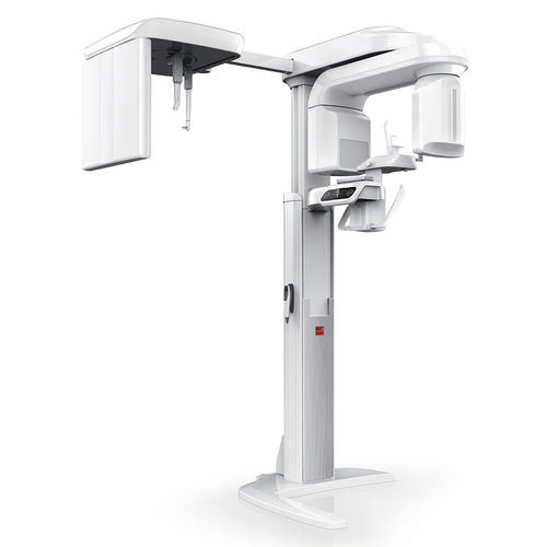
Panoramic X-ray system PaX-i3D™ cephalometric X-ray systemdental CBCT scannerdigital
Add to favorites
Compare this product
Characteristics
- Type of system
- panoramic X-ray system, cephalometric X-ray system, dental CBCT scanner
- Technology
- digital
- FOV (cm)
- 5x5 cm, 8x8 cm, 5x8 cm
- Voxel size (μm3)
120 µm, 200 µm, 300 µm
Description
Special Software for each specialty -
Analyze Ez3D-i images with advanced tools and functions
Ez3D-i supports effective and efficient communication with your patients
Wide Range of Ceph Modes -
Scan Type: LAT / Full LAT
One Shot Type: Small / Medium / Large
POWERFUL DIAGNOSTIC VALUE WITH 3D IMAGES
By selecting the appropriate FOV size, you can have the optimum image for your diagnostic needs reducing unnecessary X-ray radiation for patients.
5X5 images are useful for specific area diagnosis with minimum X-ray exposure for patients, It can especially increase the accuracy of endodontic diagnosis by exactly checking the amount or root canals and abnormal root canal shapes such as C-shapes that are difficult to check using 2D X-ray system.
8X5 images can provide more extended oral information on maxillary or mandibular areas. An accurate treatment plan can be established by taking into account the major anatomical structures like mandibular nerve, mental foramen or maxillary sinus.
8X8 images enable comprehensive diagnosis and treatment planning including both maxillary and mandibular areas in a single scan. It is useful for complex implant surgery as well \as left or right TMJ diagnosis.
12X9 images can provide the most optimal information for oral diagnosis fully covering both maxillary and mandibular structures including the 3rd molar region in a single scan. It is suitable for most oral surgery cases as well as multiple implant surgery.
Related Searches
- Analysis medical software
- Radiology software
- Tablet PC software
- Flat panel sensor
- Scheduling software
- Diagnostic medical software
- Dental software
- Dental radiography system
- Treatment software
- Tracking software
- Digital dental radiography system
- Education software
- Intraoral X-ray system
- Simulation software
- Data analysis software
- Image analysis software
- Panoramic X-ray system
- Dental CBCT scanner
- Dental X-ray scanner dental radiography
- 60 kV dental radiography
*Prices are pre-tax. They exclude delivery charges and customs duties and do not include additional charges for installation or activation options. Prices are indicative only and may vary by country, with changes to the cost of raw materials and exchange rates.


