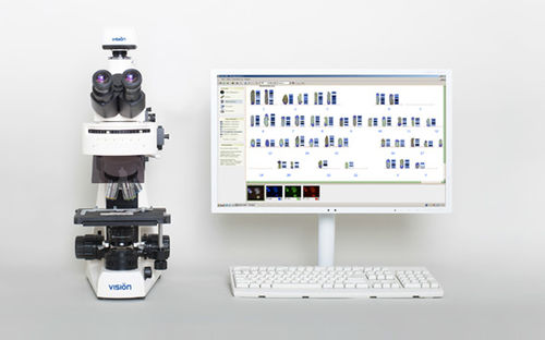
Automatic cell imaging system Vision Karyo FISHfor karyotypinglaboratoryfor DNA
Add to favorites
Compare this product
Characteristics
- Operation
- automatic
- Applications
- for karyotyping, laboratory
- Cell type
- for DNA
- Observation technique
- FISH
Description
Karyotyping and analysis using the FISH method
Automatic karyotyping of chromosomes
A modern approach to chromosome analysis, using FISH method
Digital camera
High resolution delivers superior quality image of a metaphase plate. An ultrasensitive camera detects even the weakest signals.
Toolbar
The toolbar is designed according to the analysis’ algorithm and ensures compliance with all the stages of the procedure, providing reliable results.
Optical system
The combination of innovative technology and classical microscopy extends the working possibilities. If necessary, microscopy sample of a metaphase plate can be viewed through the eyepieces.
Karyotyping
An automated karyotyping with the possibility of manual correction.
Fluorescence
A fluorescent unit with up to 6 filters provides a wide range of possibilities for the FISH method application.
Final image and pseudocoloring
The final image is generated by combining and pseudocoloring a serie of original monochrome images with different fluorescent stains.
High throughput and quality
Die automatische Karyotypisierung von Chromosomen
Vision Karyo automatically detects changes in the number and structure of chromosomes, which allows you to diagnose genetic abnormalities(Down, Patau, Edwards and other syndromes).
Vision Karyo automatically detects changes in the number and structure of chromosomes, which allows you to diagnose genetic abnormalities(Down, Patau, Edwards and other syndromes).
Karyotyping of plant and animal chromosomes
Karyotyping of plant and animal chromosomes
Vision Karyo allows the control of genetic material structure in breeding new varieties of plants and animals.
Catalogs
No catalogs are available for this product.
See all of West Medica‘s catalogsRelated Searches
- Automated cell imaging system
- Cell imager
- Laboratory cell imager
- Fluorescence cell imaging system
- Diagnostic cell imaging system
- Molecular biology cell imaging system
- Phase contrast cell imaging system
- Bright field cell imaging system
- Blood cell cell imaging system
- Pathology cell imaging system
- Clinical cell imaging system
- Veterinary laboratory cell imaging system
- Hematology cell imaging system
- FISH cell imaging system
*Prices are pre-tax. They exclude delivery charges and customs duties and do not include additional charges for installation or activation options. Prices are indicative only and may vary by country, with changes to the cost of raw materials and exchange rates.

