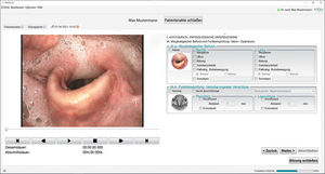
Medical software DiVAS for endoscopyoperating roomanalysis
Add to favorites
Compare this product
Characteristics
- Applications
- medical, for endoscopy, operating room
- Function
- analysis, visualization
Description
Two modules are available in DiVAS for comprehensive image documentation in 2D and 3D, the DiVAS Photo Documentation module and the DiVAS Video Documentation module.
During an examination, photos and videos can be taken and automatically assigned to the selected patient. Of course, existing image material can also be imported into a patient file at any time.
The resulting image material can be viewed and edited, for example it is possible to crop individual areas of an examination video for closer examination. In addition, there are many other functions available including exporting or importing image material or adjusting the image contrast or brightness, or changing the playback speed of a video.
Besides the image material generated with XION's own systems, image material from external signal sources such as operating room microscopes, ultrasound or fluoroscopy can also be integrated into DiVAS for further documentation and processing.
Related Searches
- Analysis software
- Viewer software
- Medical imaging monitor
- Diagnostic software
- Surgical monitor
- Endoscopy monitor
- Evaluation software
- 27" monitor
- 24" monitor
- Diagnostic display
- 32" monitor
- 21.5" monitor
- 55" monitor
- Fanless monitor
- Surgery unit software
- Operating room software
- Endoscopy software
- HD video recorder
- 22" monitor
- 23.8" monitor
*Prices are pre-tax. They exclude delivery charges and customs duties and do not include additional charges for installation or activation options. Prices are indicative only and may vary by country, with changes to the cost of raw materials and exchange rates.


